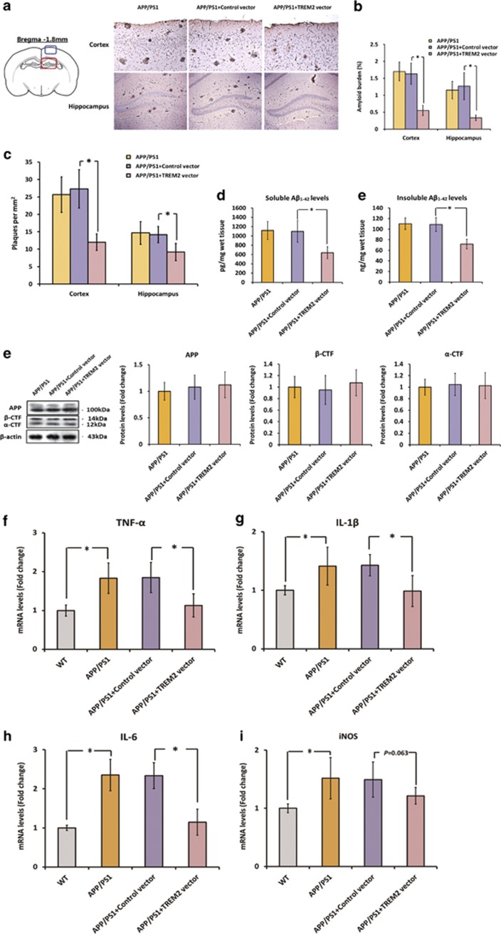
Figure 4.
Triggering receptor expressed on myeloid cells 2 (TREM2) overexpression ameliorates amyloid-β (Aβ) deposition and neuroinflammation in the brain of APPswe/PS1dE9 mice. First, a lentiviral strategy was used to overexpress TREM2 in the brain of APPswe/PS1dE9 mice. (a) Represent photos of amyloid plaques in the cortex and hippocampus of APPswe/PS1dE9 mice injected with vehicle, control vector or TREM2 vector. Amyloid plaques were detected by immunohistochemistry using an anti-Aβ (4G8, which recognizes amino-acid residues 17–24 of Aβ) antibody. The plaques were visualized by a microscopy with × 200 magnification; in the cortex: scale bar=100 μm; in the hippocampus: scale bar=200 μm. Blue box on the left atlas shows the region where the photo of the cortex was taken, and red box on the left atlas shows the region where the photo of the hippocampus was taken. (b) The amyloid burden in the cerebral cortex and hippocampus was expressed as the percentage of the area reactive with anti-Aβ antibody (4G8) in relation to the total area analyzed. (c) The amyloid plaque density (number of plaques per mm2) was also calculated in the cerebral cortex and hippocampus. (d) The levels of soluble and insoluble Aβ1–42 in the brain were detected by enzyme-linked immunoassay (ELISA). (e) The protein levels of full-length APP, β-CTFs, and α-CTFs in the brain were measured by western blot analysis. Data of western blot analysis were normalized to β-actin. It should be noted that cropped gels are used here, and the full-length gels are shown in Supplementary Figure S6. (f–i) mRNA levels of TNF-α, IL-1β, IL-6, and iNOS in the brain of APPswe/PS1dE9 mice injected with vehicle, control vector, or TREM2 vector. All data were analyzed by one-way analysis of variance (ANOVA) followed by Tukey's post hoc test. Columns represent mean±SEM (n=8 per group). *P<0.05. APP, amyloid precursor protein; β-CTFs, C-terminal fragment cleaved by β-secretase; α-CTFs, C-terminal fragment cleaved by α-secretase.

