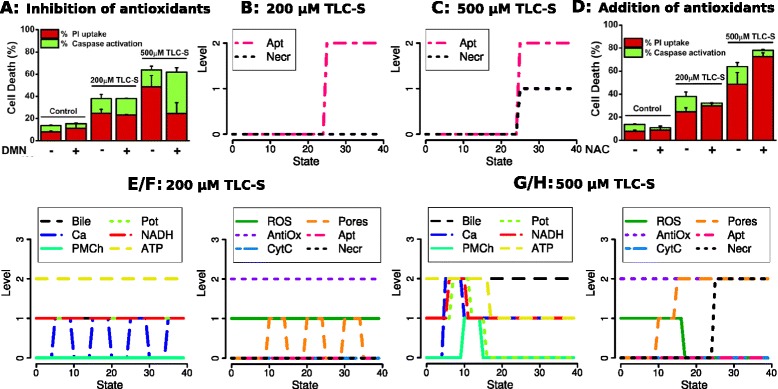
Figure 5.

Data (bar plots) and simulations for the inhibition and addition of antioxidants. A: Fraction of apoptotic and necrotic cells with inhibition of the antioxidant NQO1 by DMN. B : Simulations during 200 μM bile (TLC-S) stimulation, C : Simulations during 500 μM TLC-S stimulation. For this condition, data and simulations exhibit increased apoptosis together with decreased necrosis (cf. Figure 2). The same enhancement of apoptosis is predicted already for 200 μM, whereas few changes are actually measured. D – H (addition of the antioxidant N-acetyl-l-cysteine (NAC)): In comparison to Figure 2, apoptosis decreases to level 0 for both stimulating conditions, but necrosis remains unchanged (0/2). In the simulations, NADH does not decrease. In contrast to Figure 2C, for 500 μM TLC-S stimulation ROS decrease to 0 after strong pore opening and potential breakdown. Figures A and D are reprinted from ([2], Figure four C and B), with permission from Elsevier.
