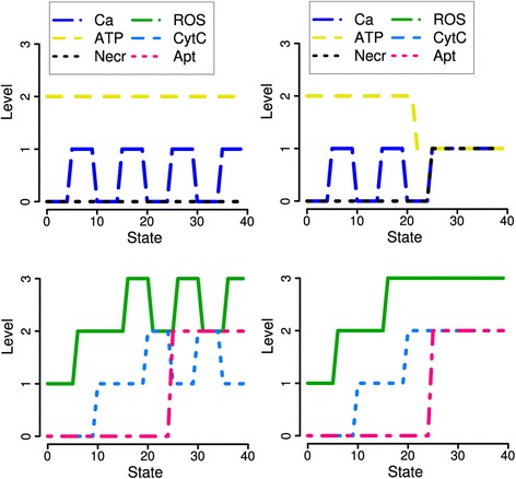
Figure 6.

Simulations for 200 μM TLC-S stimulation of acinar ( left ) and liver cells ( right ). Inhibition of antioxidants enables ROS increase up to the maximal level 3. Liver cells are reported to be more sensitive to ROS induced mitochondrial depolarization, pore opening, ATP depletion and subsequent necrosis [1]. Thus, ROS have a role similar to the role of Ca2+ in acinar cells stimulated with 500 μM TLC-S (cf. Figure 2).
