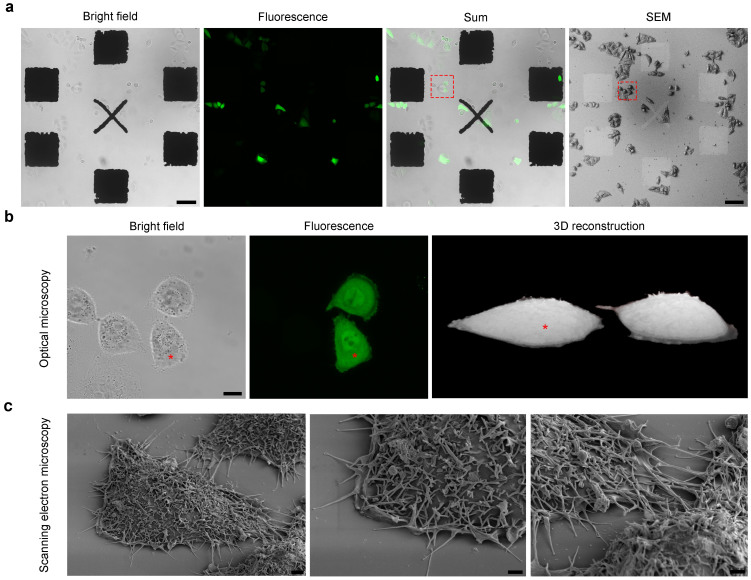
Figure 3. High-resolution optical and electron microscopy images of HeLa cells expressing EGFP.
(a) Fields of view showing HeLa cells overexpressing soluble EGFP growing on the patterned coverslip as acquired by means of optical microscopy (Bright field, Fluorescence and Sum) and SEM. Scale bars 100 μm. The pattern was developed so that it could be easily visualised by both microscopes in order to allow the rapid relocation of the same cells when moving from one to the other. (b) High-resolution optical images (Bright field and Fluorescence maximum projections), and a 3D reconstruction of the two EGFP-expressing cells (red box) in (a). Scale bar: 10 μm. (c) Scanning electron microscopy images of the EGFP-expressing HeLa cell asterisked in (b) showing the highly preserved ultrastructure obtained after SEM preparation on the patterned substrates, and the high-resolution details observable after tilting the sample holder. Scale bars 2 μm.

