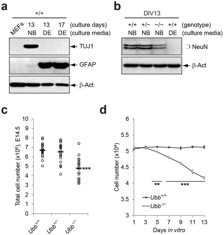
Figure 1. Reduced number of Ubb−/− cells during culture in vitro.
(a) Immunoblot detection of neuronal marker TUJ1 and glial cell marker GFAP in cells isolated from wild-type (+/+) embryonic brains on 14.5 dpc that were cultured under two different medium conditions. Growth of neurons was facilitated in Neurobasal® medium with B-27 supplement (NB), whereas growth of glial cells was preferred in DMEM/10% FBS (DE). β-Actin (β-Act) was used as a loading control, and mouse embryonic fibroblasts (MEFs) were included as a negative control. (b) Immunoblot detection of neuronal marker NeuN at 48 and 46 kDa in cells isolated from wild-type (+/+), Ubb+/− (+/−), and Ubb−/− (−/−) embryonic brains on 14.5 dpc and cultured in vitro for 13 days (DIV13). Wild-type (+/+) cells were cultured in two different medium conditions (NB and DE). (c) Determination of total cell numbers in Ubb+/+ (n = 19), Ubb+/− (n = 25), and Ubb−/− (n = 17) embryonic brains on 14.5 dpc. (d) Cells isolated from Ubb+/+ (n = 4) and Ubb−/− (n = 3) embryonic brains were plated on 96-well plate at 5 × 10⁴ cells/well and cultured in vitro and counted using a hematocytometer. Representative immunoblot results of cells from two different embryonic brains per genotype are shown (a, b), and data are expressed as the means ± SEM from the indicated number of samples (c, d). **P < 0.01; ***P < 0.001 vs. Ubb+/+ (c) or Ubb+/+ on each day (d).

