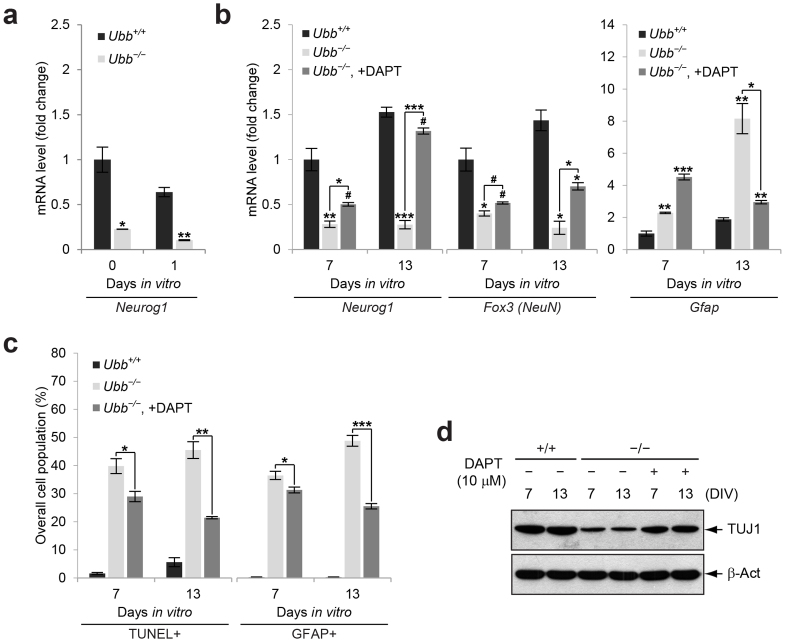
Figure 5. Improvement of Ubb−/− neuronal and glial phenotypes via pharmacological intervention of Notch signaling.
(a) Expression levels of the proneuronal gene Neurog1 in wild-type (Ubb+/+) and Ubb−/− cells (n = 3 each) on DIV0 and DIV1 were determined by qRT-PCR as described in Figure 4(a). (b) Expression levels of Neurog1, Fox3 (NeuN), and Gfap in wild-type (Ubb+/+) and Ubb−/− cells (n = 3 each) on DIV7 and DIV13 were determined by qRT-PCR as described in Figure 4(b). DAPT treatment of Ubb−/− cells was carried out as described in Figure 4(b). (c) The percentage of apoptotic (TUNEL+) or GFAP+ cells in wild-type (Ubb+/+), Ubb−/−, and DAPT-treated Ubb−/− cells (n = 3 each) on DIV7 and DIV13 was determined in a similar manner as described in Figure 2(a). (d) Immunoblot detection of TUJ1 in wild-type (Ubb+/+), Ubb−/−, and DAPT-treated Ubb−/− cells on DIV7 and DIV13. β-actin (β-Act) was used as a loading control. Representative immunoblot results of cells from three different embryonic brains per genotype are shown (d), and data are expressed as the means ± SEM from the indicated number of samples (a–c). #P < 0.1; *P < 0.05; **P < 0.01; ***P < 0.001 vs. Ubb+/+ on each day or as indicated by bars.

