. 2014 Nov 13;3:e03895. doi: 10.7554/eLife.03895

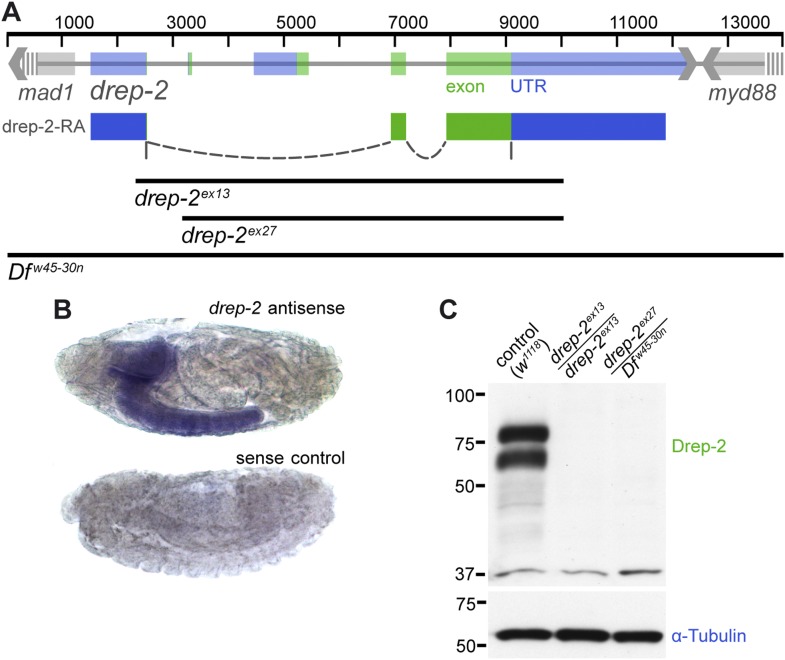
Figure 1. Expression and mutants of drep-2.

(A) Genetic scheme of the drep-2 locus on chromosome IIR. The neighboring genes mad-1 and myd88 extend beyond the sequence displayed. The cDNA labeled drep-2-RA was used for rescue experiments. Blue: untranslated regions; green: exons; black lines: deleted regions in the mutants. (B) In situ hybridization of drep-2 reveals a neuronal expression pattern (stage 17). (C) Western blot of adult fly head extracts using the anti-Drep-2C-Term antibody. Drep-2 isoforms are predicted to run at 52 and 58 kDa. The signal is absent in both the drep-2ex13 and the drep-2ex27/Dfw45-30n mutant.

DOI: http://dx.doi.org/10.7554/eLife.03895.003

Figure 1.

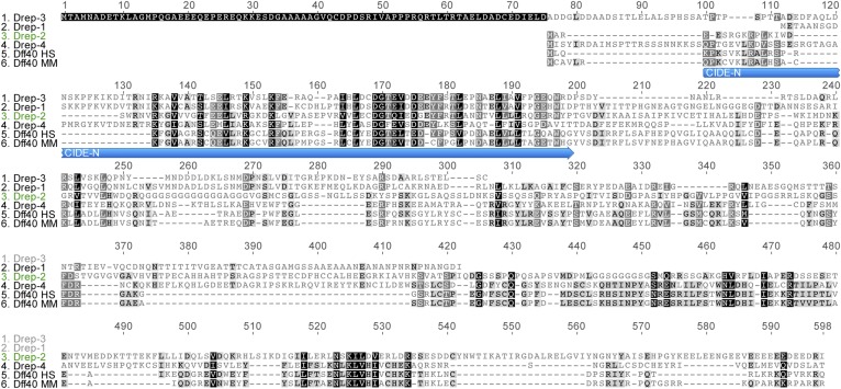
Figure 1—figure supplement 1. Drep protein alignment.

Figure 1—figure supplement 1.

Sequence alignment of all four Drosophila Dff proteins, as well as human (HS) and murine (MM) Dff40. Drep-4 has the strongest similarity to Dff40, yet Drep-2 also shows conserved motifs in addition to the CIDE-N domain. The alignment was created using Geneious v5.3.6. (http://www.geneious.com)
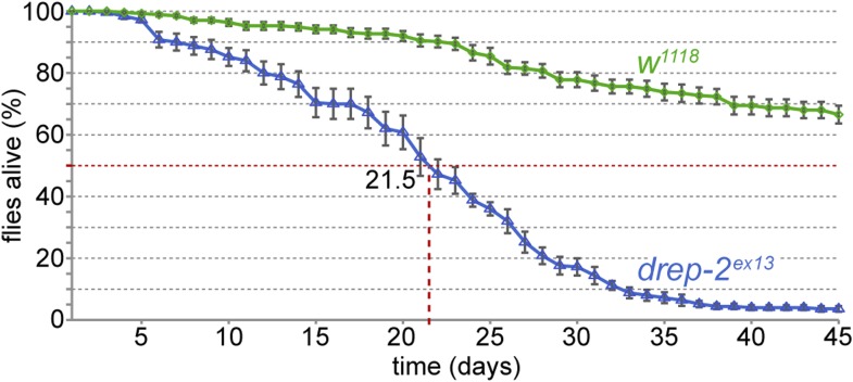
Figure 1—figure supplement 2. Reduced lifespan of drep-2ex13 mutants.

Figure 1—figure supplement 2.

Comparison to isogenic w1118 control flies: 50% of mutant flies were dead after 21.5 days. Mutant: n = 10 vials (each containing 25 flies), control: n = 11.