. 2014 Nov 13;3:e03895. doi: 10.7554/eLife.03895

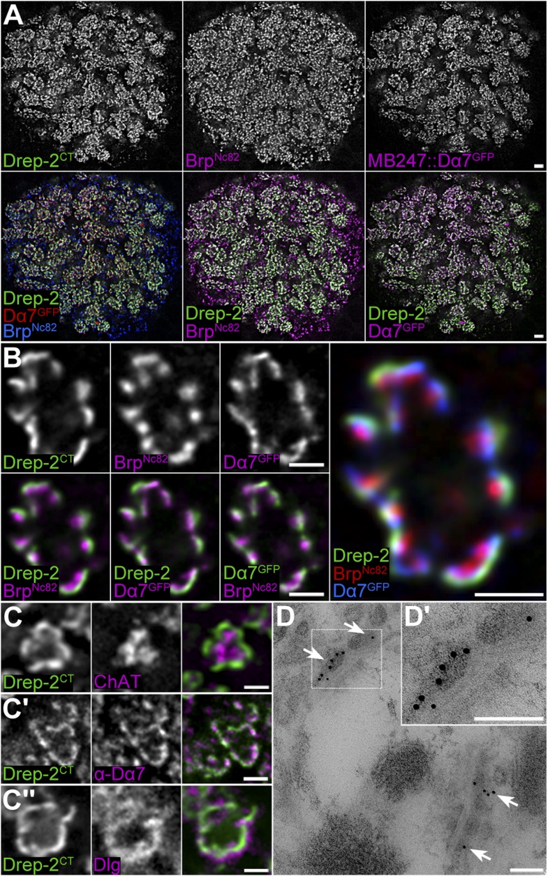
Figure 4. Drep-2 is enriched at KC postsynapses.

(AB) Drep-2C-Term and BrpNc82 staining in animals expressing the construct mb247::Dα7GFP that marks acetylcholine receptors in MB KCs. (A) Detailed image of the MB calyx. Scale bar: 2 µm. (B) Detail of a single microglomerulus in the calyx. Drep-2C-Term overlaps with postsynaptic mb247::Dα7GFP and not with presynaptic Brp. Scale bars: 1 µm. (C) Localization of Drep-2 relative to choline acetyltransferase (ChAT, presynaptic cytosol, C), the postsynaptic ACh receptor subunit Dα7 (antibody staining, C′), and the postsynaptic scaffolding protein Discs large (Dlg, C″). Drep-2 colocalizes with postsynaptic markers. Scale bars: 1 µm. (D) Post-embedding immunoelectron microscopy of Drep-2C-Term in the calyx. Arrows: Clusters of postsynaptic Drep-2C-Term. Scale bars: 100 nm.

DOI: http://dx.doi.org/10.7554/eLife.03895.008

Figure 4.

Figure 4—figure supplement 1. Drep-2 localizes to postsynaptic membranes of KCs in the calyx.

Figure 4—figure supplement 1.

(A) STED microscopy superresolution recording of Drep-2C-Term; the BrpNc82 channel is in normal confocal mode. The Drep-2 signal does not overlap with presynaptic Brp. Scale bar: 1 µm. (BE) Expression of drep-2 constructs in KCs yields a label resembling the Drep-2 antibody staining. Comparison to BrpNc82; all scale bars: 1 µm. (B) Pan-neural overexpression. Elavc155-Gal4 and UAS-Drep-2mStrawberry; mStrawberry signal is shown. (C) KC-specific overexpression. C305a-Gal4, UAS-Drep-2mStrawberry, and UAS-Dα7GFP; mStrawberry and GFP signals are shown. D-2 = Drep-2mStrawberry, D7 = Dα7GFP. (D) PN-specific overexpression. Gh146-Gal4 and UAS-Drep-2mStrawberry; diffuse mStrawberry is shown. (E) KC-specific expression of UAS-Drep-2 in the drep-2ex13-mutant background. Mb247-Gal4 and untagged UAS-Drep-2; Drep-2C-Term staining is shown.