Figure 3.

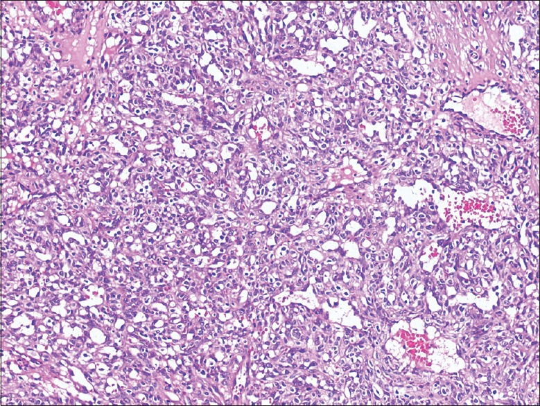
H and E stained micropictograph shows ulcerated stratified squamous keratinized epithelium with underlying granulation tissue. Numerous varied caliber of blood vessels arranged in lobular pattern is seen in the connective tissue

H and E stained micropictograph shows ulcerated stratified squamous keratinized epithelium with underlying granulation tissue. Numerous varied caliber of blood vessels arranged in lobular pattern is seen in the connective tissue