Figure 4.

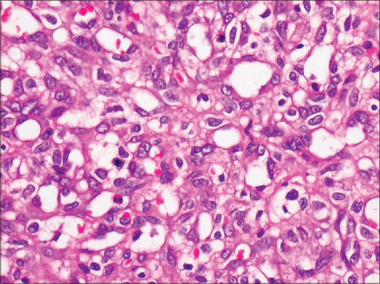
H and E stained section shows numerous blood vessels of varying size lined by endothelial cells. Few dilated blood vessels are also seen (×40). There is endothelial cell proliferation admixed with mixed inflammatory cell infiltrate

H and E stained section shows numerous blood vessels of varying size lined by endothelial cells. Few dilated blood vessels are also seen (×40). There is endothelial cell proliferation admixed with mixed inflammatory cell infiltrate