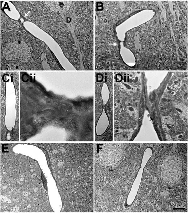
Figure 11.

Microvascular strictures in the blast-exposed brain. Longitudinal sections of microvessels from animals that were exposed to either one (A, B) or three (C, D) 74.5 kPa blasts and were sacrificed 24 hours later. Strictures where there is narrowing of the vascular lumen are indicated by arrows. The dendrite (D) of a nearby neuron is indicated in panel A. Panel Ci shows a region exhibiting a microvascular stricture (box in Ci). A higher power image shows that the lumen of this microvessel has been occluded by amorphous material and opposing endothelial cell walls appear to have fused (Cii). Panel D shows complete luminal occlusion by amorphous material. The boxed region in panel Di is illustrated at higher power in panel Dii. Note that despite the destruction of the microvessel architecture at the site of the strictures, the surrounding neuropil appears normal. Panels E and F illustrate longitudinally cut microvessels from non-blast exposed control brains. Scale bar 1 μm A-B; 1.2 μm Ci; 0.2 μm Cii; 2.5 μm Di; 0.5 μm Dii; 3.5 μm E-F.
