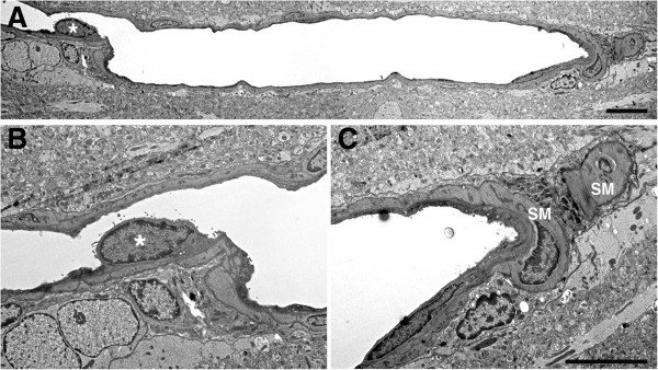
Figure 14.

Blast-exposed microvessel with degenerative changes. A longitudinal section of a microvessel is shown from an animal that received a single 74.5 kPa blast exposure and was harvested 24 hours later. An endothelial cell nucleus that has been displaced into the vascular lumen is indicated by an asterisk in panels A and B. At the opposite end of the microvessel the smooth muscle (SM) layers are disrupted. The microvessel lumen is also irregular. Scale bars: 1.5 μm A; 3 μm B, C.
