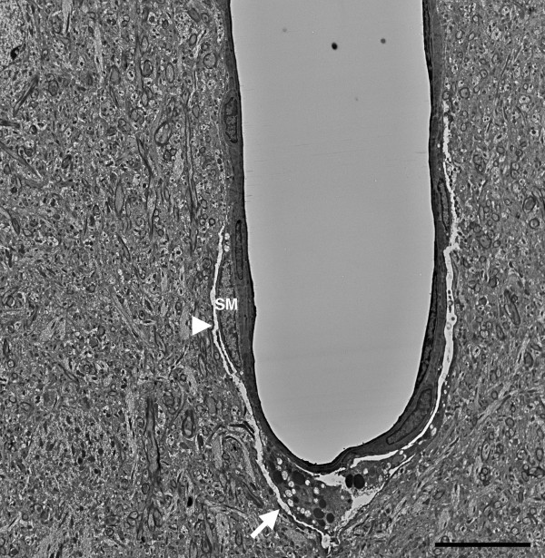
Figure 17.

Chronic pathology in a penetrating cortical vessel following blast exposure. Transverse section of a cortical penetrating vessel from the frontal cortex of a rat that received three 74.5 kPa blast exposures and was sacrificed 6 months after the last exposure. Note the disruption of the tunica media at the site of the arrow and the presence of a vacuolated region (arrowhead) that extends into the adventitia. A smooth muscle (SM) cell is indicated. The surrounding neuropil appears normal. Scale bar: 10 μm.
