Figure 1.
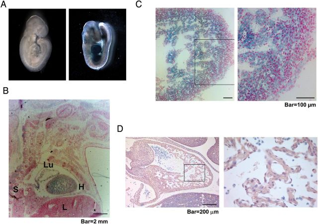
β2-Spectrin is prominent in the developing heart. (A) X-gal staining of a whole-mount Spnb2+/tm1a embryo (right) at E9.5 revealed the expression of β2-spectrin. (B) X-gal staining of a frozen section of a Spnb2+/tm1a embryo at E16.5. H, heart; L, liver; Lu, lung; S, spinal cord. (C) At E16.5, expression of β2-spectrin was detected in both compact and trabecular areas of the heart. (D) Normal heart from a wild-type embryo at E12.5 was stained with the antibody against β2-spectrin. The right panels represent magnifications of the boxed area. Scale bars, 2 mm (B); 100 μm (C); 200 μm (D).

