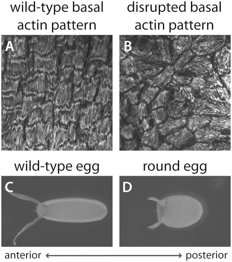
Fig. 2.
Relationship between the basal actin pattern and final egg shape. (A–B) Organization of contractile actin bundles at the basal surface of the follicular epithelium at stage 12. The F-actin is visualized with rhodamine phalloidin. (A) In a wild-type epithelium, the basal actin bundles are oriented perpendicular to the AP axis, which creates a circumferential pattern around the egg chamber. (B) Under certain mutant conditions, the basal actin bundles are still largely aligned within individual cells, but their tissue-level organization is lost. (C–D) Dorsal views of mature eggs. (C) A wild-type, elongated egg. (D) The rounded egg shape that arises from the disrupted tissue pattern shown in (B).

