
Fig.1. Differences in mouse muscle microtubule organization from wt to mdx mice can be difficult to assess visually.
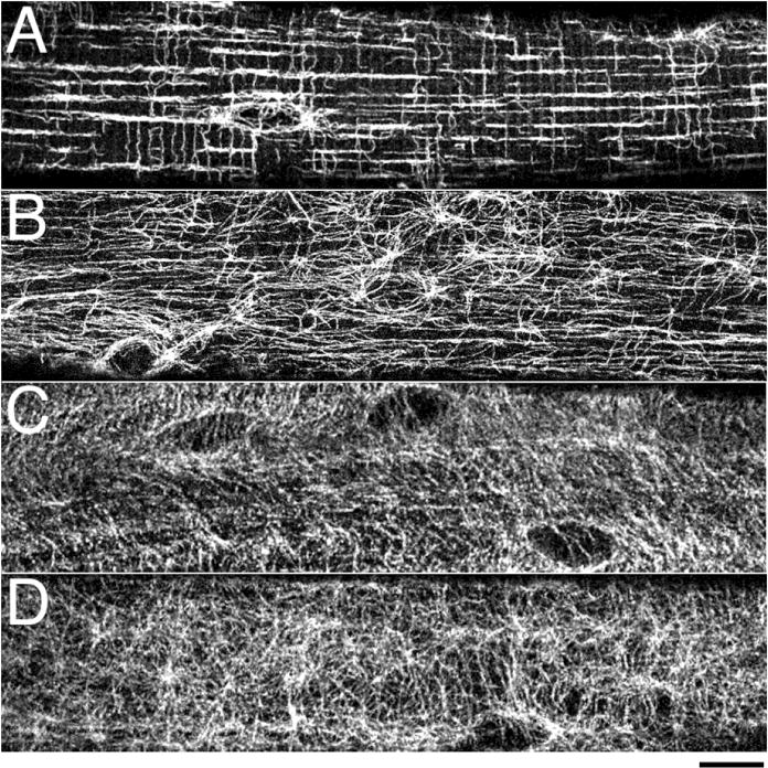
Single muscle fibers were stained with anti-α-tubulin: fast-twitch EDL muscle fibers from wt (A) and mdx mice (B, the mouse model for Duchenne muscular dystrophy); slow-twitch SOL muscle fibers from wt (C) and mdx (D) mice. EDL wt fibers show an organized microtubule network with longitudinal and transverse components, the loss of which in mdx is easy to notice visually. SOL wt fibers have a less regular network; differences between wt and mdx are difficult to assess. Each panel shows a single confocal image focused on the cortical layer of microtubules that surround the fiber core. Bar: 10μm.