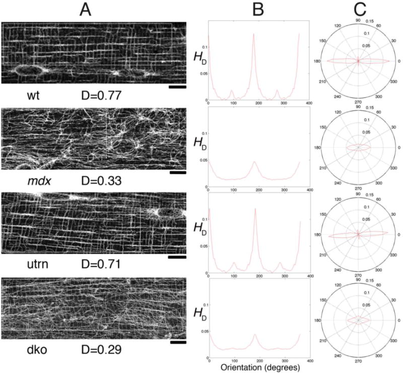
Fig. 4. Microtubule directionality detection by TeDT in fast-twitch muscle fibers from different mouse genotypes.

TeDT was used to compare microtubule organization in fibers from the fast-twitch EDL muscle of wild-type (wt), mdx, utrn (in which dystrophin’s homologue utrophin is knocked-out) and mdx-utrn double knockout (dko) mice. The left column shows a confocal image for each mouse group. The analysis was carried out on the regions of interest drawn with white dots to exclude the nuclei (arrows) which have their own organization. The directional histograms (HD) from 0 to 360° are presented in Cartesian (central column) and in polar coordinates (right column). D is the directionality score (see Methods) for each of the images. Bar: 10μm.
