
Fig. 6. Microtubule directionality detection by TeDT in slow-twitch muscle fibers from different mouse genotypes.
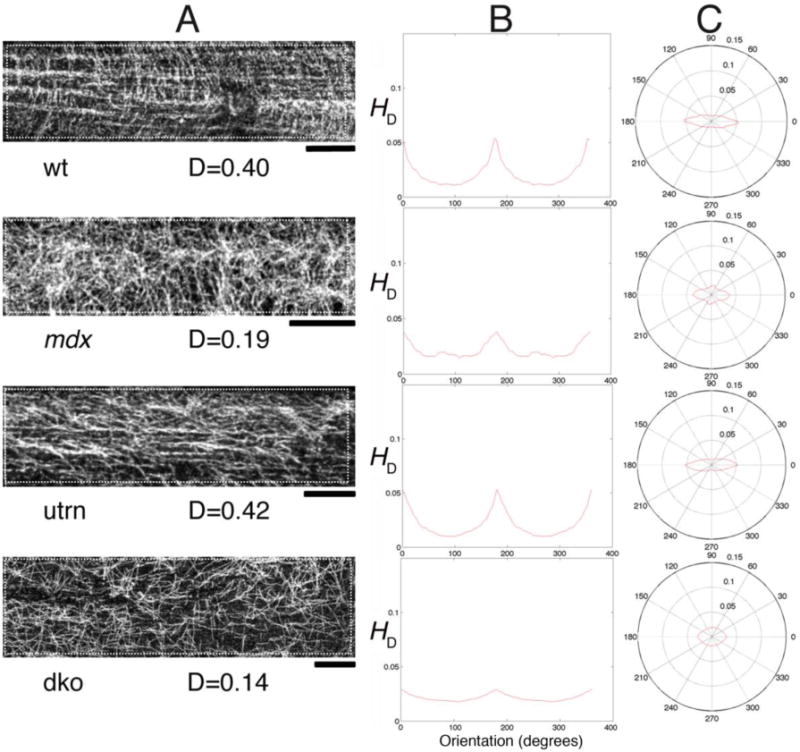
Microtubules of SOL muscle fibers from wt, mdx, utrn and dko mice were analyzed as those of EDL were in Fig. 4. The left column shows a confocal image for each mouse group. The directional histograms (HD) from 0 to 360° in Cartesian and in polar coordinates are shown in the middle and right columns respectively. Bar: 10μm