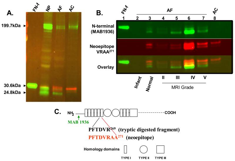
Figure 2. The fibronectin (FN) antibody to neoepitope VRAA271 is specific for the fibronectin fragment (FN-f), while the N-terminal antibody (mAB 1936) binds to both full length fibronectin and its fragments.
Panel A: Western blot using the neoepitope VRAA271 antibody (shown in red) and the N-terminal antibody (mAB 1936, shown in green). Lanes, from left to right: 29-kDa fibronectin fragment (FN-f); degenerative nucleus pulposus (NP) tissue; degenerative human annulus fibrosus (AF) tissue; arthritic articular cartilage (AC, as positive control). Panel B: FN and FN-f species in human AF tissues by western blot. Lane 1: human plasma derived 29-kDa FN-f; lane 2: infant AF tissue; lane 3: normal young adult AF tissue; lane 4-7: adult AF surgical samples with increasing degree of degeneration; Lane 8: arthritic AC. Panel C: Schematic diagram of the human FN protein structure. Arrows below the diagram illustrate the location of the antibody epitopes; Diagram modified from Hynes (1990);3 numbering of amino acids according to Zack et al. (2009).16

