
Figure 5.
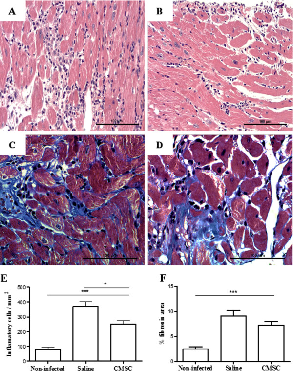
Morphometric analysis of heart sections from uninfected and chagasic mice treated with cardiac mesenchymal stem cells (CMSCs) or saline. Representative images of sections of hearts from mice euthanized 2 months after cell therapy with CMSCs and untreated controls are shown: heart sections of animals untreated (A) and treated (B) stained with hematoxylin and eosin (H&E) and heart sections stained with Masson’s trichrome obtained from untreated (C) and treated (D) mice. (E) Number of inflammatory cells per mm2 measured in sections stained with H&E. (F) Percentage of fibrosis quantified in sections stained with Masson’s trichrome. Results are expressed as mean ± standard error of the mean of 5 to 10 animals per group. *P <0.05. *** p < 0.001.