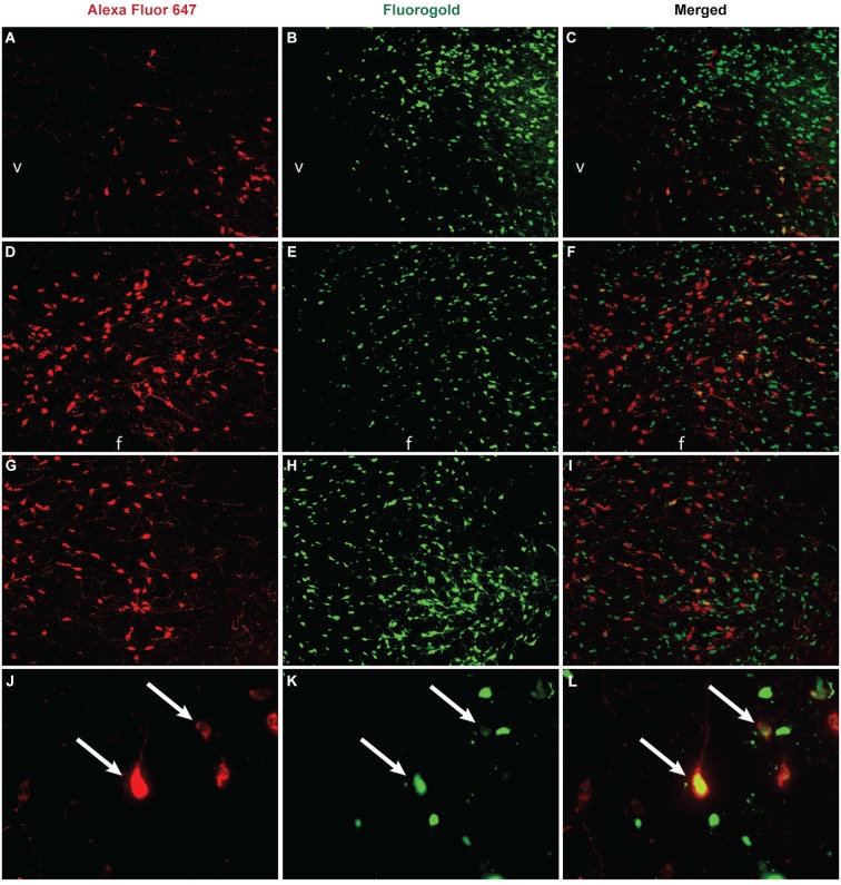
Figure 1.
Unpublished data from authors. Co-labeling between orexin and VTA projection neurons in the hypothalamus. The retrograde tracer Fluoro-Gold (Fluorochrome, Denver CO) was microinjected (2% in 250 nl) into the VTA, brains were collected and sliced at 40µm, and then tissue was stained for orexin A immunoreactivity via overnight incubation with an anti-orexin A antibody (1:8000, Phoenix Pharmaceuticals, Burlingame CA) and visualized through incubation with Alexa Fluor 647 (1:200, Jackson ImmunoResearch, West Grove PA) for 1 h. Images (A–I) were taken at 10x magnification and images (J–L) were taken at 40x. In the DMH (A–C), significant orexin neuron labeling was observed (A) in addition to retrogradely labeled neurons from the VTA (B), and some orexin neurons projected to the VTA (C, yellow labeling). In the PeF (D–F) a subset of orexin neurons (D) and retrogradely labeled neurons from the VTA (E) co-localized (F). In the LHAd (G–I) a subset of orexin neurons (G) and retrogradely neurons from the VTA (H) colocalized (I). A 40x magnification of labeling observed in the DMH is presented in panels (J–L). Double-labeled neurons are indicated by white arrows. v = ventricle, f = fornix.

