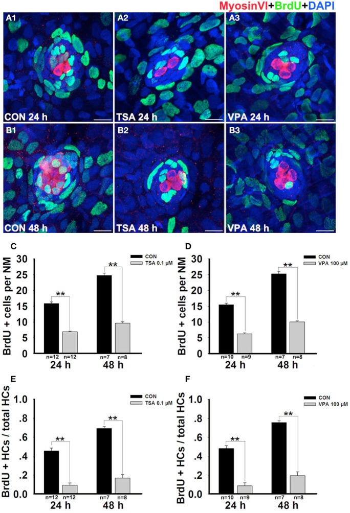
Figure 4.
HDAC inhibition decreases the proportion of cells in S-phase. (A and B) Lateral line HCs are stained with Myosin VI, and the BrdU antibody shows dividing cells in the neuromasts of zebrafish. (C and D) BrdU+ cells were counted in control and inhibitors-treated larvae at 24 h and 48 h after neomycin damage. (E and F) Quantification of the ratio of BrdU+ HCs in control and inhibitors-treated larvae at 24 h and 48 h after neomycin incubation. Bars are mean ± s.e.m. and n = total number of embryos. **p < 0.001.

