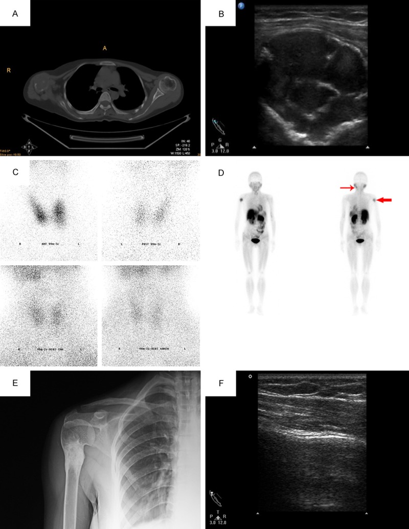
Figure 2.

A. CT demonstrated a well-circumscribed large mass in the right humeral head, with bony expansion; B. The ultrasound showed that there was a cystic-solid mass in the right humerus. The tumor was of an irregular shape, with clear margins, and hyperechoic ribbons within; C. None focal accumulation of radiotracer uptaked in the normal position of parathyroid; D. focal accumulation of radiotracer uptaked in the right upper humeral shaft (brown tumor) and the left mandible (ectopic parathyroid adenomas); E. 30 months after parathyroidectomy, X-ray revealed reactive sclerosis on the original diseased region; F. The ultrasound showed well bony cortex and the mass on the original diseased region of the right humerus was disappeared.
