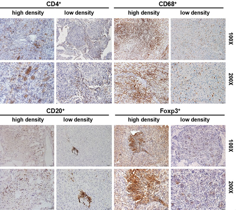
Figure 1.

CD4+ T lymphocyte subset, CD20+ B lymphocyte, CD68+ macrophages, and FOXP3+ cells in tumor tissues. Immunohistochemistry was performed by using the anti-CD4, anti-CD20, anti-CD68 and anti-Foxp3 antibodies, respectively. The low density and high density cells in the tumor tissues were shown.
