
Figure 3.
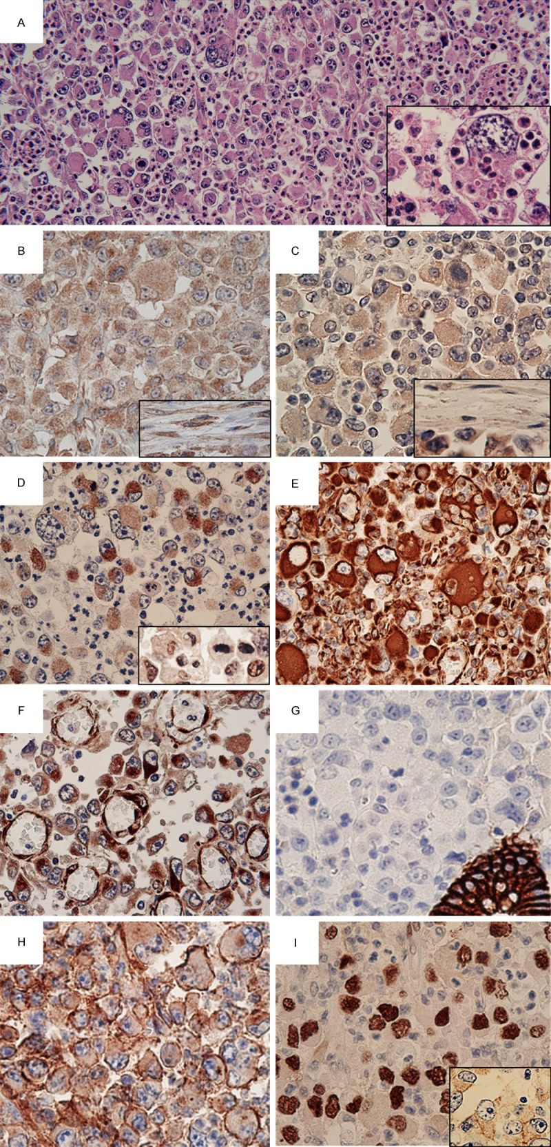
Microscopic findings with hematoxylin and eosin (HE) staining and immunohistochemistry. A. The tumor was composed of pleomorphic large and giant cells with loose or no cohesion to each other. There were abundant neutrophils surrounding the tumor (HE, ×200). Neutrophils were present within tumor cells (emperipolesis) (inset, ×600). B. Tumor cells stained positive for granulocyte colony-stimulating factor (×400). Note the positive reaction for stromal fibroblasts/myofibroblasts (inset, ×400). C. Immunoreactivity for granulocyte colony-stimulating factor receptor (G-CSFR) was observed in tumor cells (×400); stromal fibroblasts/myofibroblasts were negative for G-CSFR (inset, ×400). D. AE1/AE3 immunostaining demonstrated focal positivity in the tumor cells (×400). Inset: some tumor cells were positive for HNF4α (×400). E. Strong reactivity of the tumor cells for vimentin (×400). F. In addition to positive staining for nestin in tumor cells, endothelial cells in highly proliferating small blood vessels were highlighted via immunoreactivity for nestin (×400). G. Negativity of E-cadherin in tumor cells. Note the positive reaction to a pre-existing crypt (×400). H. Overexpression of CD44 was observed in tumor cells (×400). I. Diffuse nuclear accumulation of p53 was detected (×400). Membranous positivity of epidermal growth factor receptor was also present (inset, ×400).