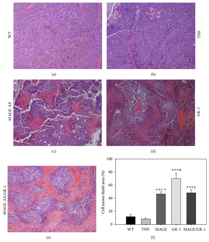
Figure 6.
Histopathological analysis. Photomicrographs of histological sections of tumors from mice vaccinated with BMDCs matured with TNFα and treated with MAGE-AX, GK-1, or MAGE-AX/GK-1. Groups MAGE-AX, GK-1, and MAGE-AX/GK-1 showed plentiful tumor cell death areas. (a) No treatment (WT). Abundant tumor cells and blood vessels were observed. (b) TNFα. The same characteristics as those observed in untreated mice were observed: abundant tumor cells and blood vessels. (c) MAGE-AX. (d) GK-1. (e) MAGE-AX/GK-1. In (c), (d), and (e), pink areas (eosinophilic), composed of dead cells, were observed in addition to purple areas (basophilic), composed of very active tumor cells. (f) A graph showing the change in the areas of cell death in tumors from mice that were treated with BMDCs and which were stimulated with TNFα, MAGE-AX, GK-1, or MAGE-AX/GK-1. *** P < 0.001, ∧ P < 0.001 MAGE versus TNF, α P < 0.0001 GK-1 versus TNFα, ɛ P < 0.001 MAGE/GK-1 versus TNF. Mean ± SEM n ≥ 3.

