
Figure 1.
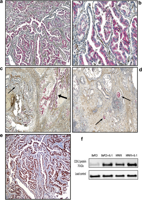
COX-2 expression in tumour tissue from pancreatic cancer. a-d Double immunostaing with monoclonal anti-COX-2 antibody (Thermo Fischer Scientific rabbit) and monoclonal anti-αSMA (Dako). COX-2 tumour positive cells (red colour), αSMA positive stromal cells (brown colour). a magnification × 100, b magnification × 200, c Heterogeneity in COX-2 expression within pancreatic cancer tissue. Areas with moderate to strong staining (thick arrow) coexist with COX-2 negative areas (thin arrow), (magnification x 100) d Moderately to strong COX-2 staining in islet cells (thin arrow), pancreatic cancer negative for COX-2 staining, (magnification x 100). e Immunohistochemistry of COX-2 expression in tumour tissue from pancreatic cancer. Immunostaining with monoclonal COX-2 mouse antibody Invitrogen (the same tumour as in a), magnification x 100. f Western blot of COX-2 expression in the moderately differentiated pancreatic cancer cell lines BxPC3 and HPAFII known to overexpress COX-2, with and without induction by interleukin 1 (Il-1), showed a specific bond for COX-2 (70 kDA) (monoclonal COX-2 mouse antibody Invitrogen).