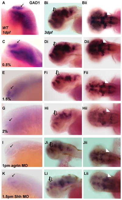
Figure 2. Effect of ethanol and MO treatment on expression of GABAergic neuronal markers.
(A, C, E, G, I and K) GAD1 expression in 1 dpf embryos. (Bi-Bii, Di-Dii, Fi-Fii, Hi-Hii, Ji-Jii and Li-Lii) GAD1 expression in 3 dpf embryos. All ethanol exposures were from 6–24 hpf. (A–Bii) WT; (C–Dii) 0.5% ethanol; (E–Fii) 1.5% ethanol; (G–Hii) 2% ethanol, (I–Jii) 1pm agrin MO and (K–Lii) 1.5pm Shh MO. The expression in 0.5% ethanol (C–Dii) is similar to that in WT embryos (A–Bii). Decreased expression is observed in all other treated embryos, for both higher concentrations of ethanol and agrin or Shh MOs (E–Lii). Arrows denote forebrain staining in 1 dpf embryos (lateral view), open arrows denote forebrain staining in 3 dpf embryos (lateral view), and arrowheads denote hindbrain staining in 3 dpf embryos (dorsal view).

