
Figure 11.
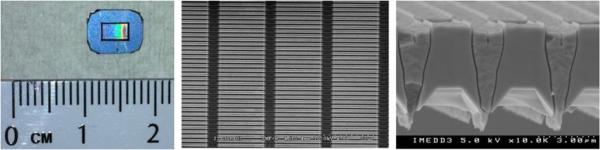
Picture of a Si nanoporous membrane showing the porous area surrounded by the support ridge (left), SEM micrographs: top view (middle) and cross-section showing the nanopores across the whole thickness of the membrane (right). Reproduced with permission from (191).