
Figure 1.
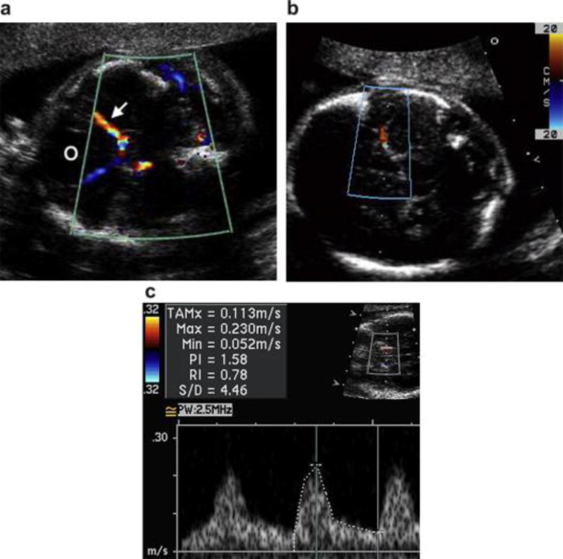
Ultrasound images in three different patients demonstrating (a) the identification of the orbits (O), circle of Willis and middle cerebral artery (MCA) (arrow) using color Doppler, (b) alignment of the pulsed wave Doppler sample volume in the proximal MCA at a low angle of insonation as it runs laterally along the petrous bone and (c) the pulsed wave Doppler spectrum and measured velocities.