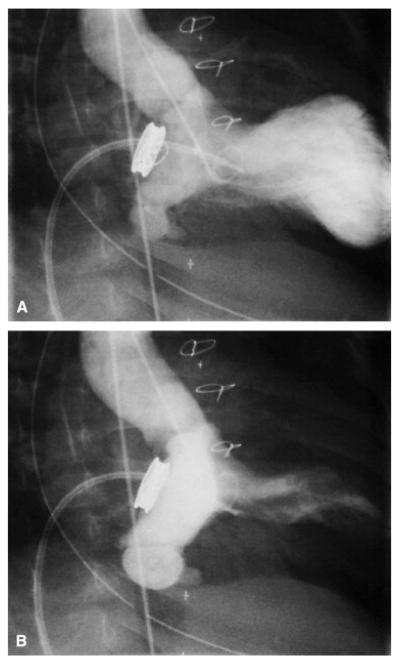
Figure E2.
Images from a retrograde left ventricular angiogram during diastole (A) and systole (B) in a patient with an aneurysm of the ventricularized portion of the left atrium. The ventricularized portion of the left atrium between the prosthesis and the native mitral valve annulus is the densely opacified area in the systolic image. The pigtail catheter was introduced into the left ventricle retrograde, through the aortic valve. The other catheter, a balloon-tipped end-hole catheter, appears to be across the mitral valve prosthesis in these images but is actually in the right ventricle.

