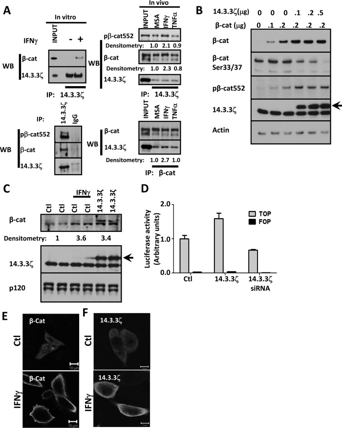
FIGURE 2:
IFNγ promotes the association of β-catenin with 14.3.3ζ. (A) Association of β-catenin with 14.3.3ζ was analyzed by coimmunoprecipitation assays. 14.3.3ζ and control immunoglobulin G (IgG) were immunoprecipitated from fresh lysates obtained from SW480 cells, control or treated with IFNγ for 1 h. 14.3.3ζ was immunoprecipitated from IECs isolated from murine intestinal mucosa exposed for 2 h to vehicle (MSA), IFNγ, and TNFα. Immunoprecipitates were blotted for β-catenin, pβ-cat552, and 14.3.3ζ. Densitometric analysis of β-catenin, pβ-cat552, and 14.3.3ζ. (B) The effect of 14.3.3ζ on β-catenin stabilization was analyzed in CHO cells. Confluent monolayers of CHO cells were transfected with 0.1–0.2 μg/ml β-catenin–expressing vector in presence of increasing concentrations of a 14.3.3ζ-expressing vector (arrow). Cell lysates were collected in RIPA lysis buffer and equal amounts of proteins loaded and analyzed by Western blotting. Actin was used as a loading control. (C) The effect of IFNγ and 14.3.3ζ (arrow) overexpression on endogenous β-catenin stability was determined by Western blot in CHO cells. Relative densitometric values were normalized with respect to the controls. p120 catenin was used as a loading control. (D) The effect of 14.3.3ζ expression on β-catenin transactivation was analyzed by TOPflash assays. SW480 cells were transfected with a vector expressing 14.3.3ζ or siRNA targeting 14.3.3ζ and luciferase expression determined. The cellular distribution of β-catenin (E) and 14.3.3ζ (F) was analyzed by immunofluorescence in SW480 cells that were exposed to vehicle (Ctl) or IFNγ for 12 h. Nuclei are blue. Bar, 10 μm.

