FIGURE 2:
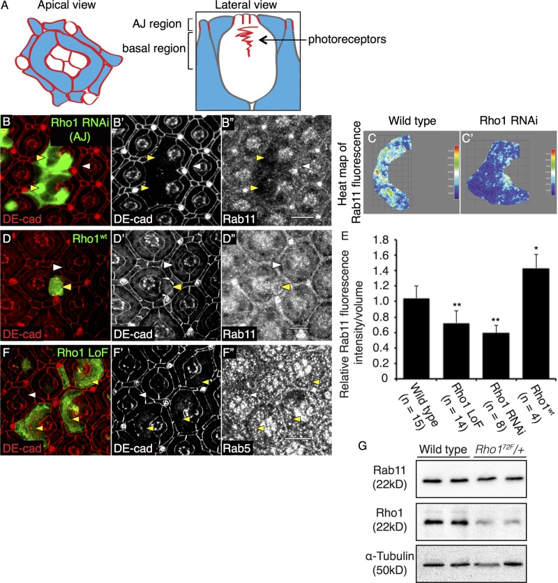
Rho1 affects staining of Rab11 recycling endosomes in the AJ region. Schematic representation of apical (left) and lateral (right) views of a pupal eye ommatidium at 41-h APF, identifying the AJ and basal regions used for analyses. Red lines represent AJ-bound DE-cadherin localization, and blue regions correspond to PECs (A). Confocal immunofluorescence localization of DE-cadherin (DE-cad, red; B, B′) and Rab11 (B′′) in AJ region of wild-type PEC clones (white arrowhead) and GFP-labeled Rho1 RNAi PEC clones (yellow arrowhead). Heat maps of sum of multiple confocal slices through the AJ region of representative PECs depicting Rab11 immunofluorescence in representative wild-type (C) and Rho1 RNAi PEC clones (C′). Confocal immunofluorescence localization of DE-cad (red; D, D′) and Rab11 (D′′) in AJ region of PECs of wild-type PEC (white arrowhead) and PEC clones overexpressing Rho1 (Rho1wt) (yellow arrowhead). Quantitation of the relative Rab11 fluorescence intensity per volume measurement in wild type, Rho172F MARCM PEC clones, and PEC clones depleted of Rho1 (Rho1 RNAi) and overexpressing Rho1 (Rho1wt) (E). The p values were calculated using an unpaired, two-sided Student's t test against values for wild-type PECs. *p < 0.05, **p < 0.01. Confocal immunofluorescence localization of DE-cad (red; F and F′) and Rab5 (endocytic vesicle marker; F′′) in wild-type PEC (white arrowhead) and GFP-labeled Rho172F MARCM clones (yellow arrowhead). Images were compiled as a sum of multiple confocal slices within the region where AJs were present, unless otherwise specified. White scale bars (lower right corner), 10 μm. Western blot analysis for Rab11 and Rho1 expression in w1118 (wild type) and Rho172F heterozygote (Rho172F/+) embryonic lysates. α-Tubulin was used as a loading control (G).

