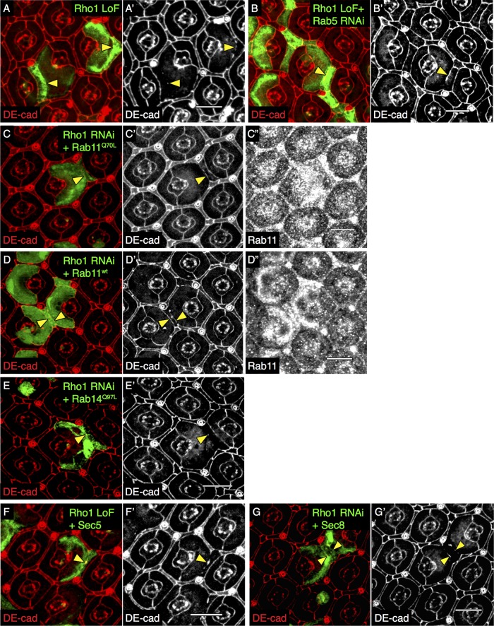
FIGURE 3:
AJ defect resulting from Rho1 loss is partially restored by Rab11 overexpression. Confocal immunofluorescence localization of DE-cadherin (DE-cad, red) in Rho172F (Rho1 LoF) MARCM PEC clones in the AJ region marked by GFP (A, A′). Confocal immunofluorescence localization of DE-cad (red) in GFP-labeled Rho172F MARCM PEC clones coexpressing Rab5 RNAi (B, B′). Confocal immunofluorescence localization of DE-cad (red; C, C′, D, D′) and Rab11 (C′′, D′′) in GFP-labeled Rho1 RNAi PEC clones coexpressing Rab11Q70L (constitutively active Rab11; C, C′′) and Rab11wt (wild-type Rab11; D, D′′). Confocal immunofluorescence localization of DE-cad (red) in GFP-labeled PECs coexpressing Rho1 RNAi and Rab14Q97L (constitutively active Rab14; E, E′), Rho172F MARCM PEC clones coexpressing Sec5 (F, F′), and Sec8 (G, G′). All images were compiled as a sum of multiple confocal slices within the region where AJs were present. Yellow arrowheads indicate disrupted (A, E–G) and rescued (B–D) AJs. White scale bars (lower right corner), 10 μm.

