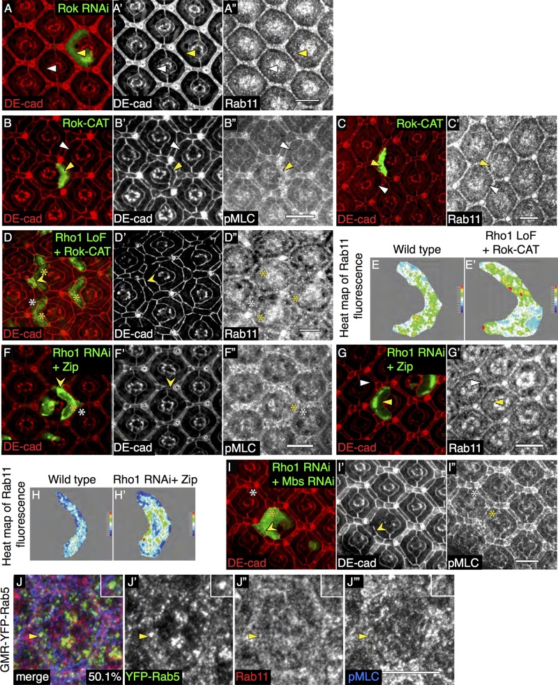
FIGURE 5:
Regulators of myosin II downstream of Rho1 affect AJ remodeling and Rab11 staining. Confocal immunofluorescence localization of DE-cadherin (DE-cad, red; A, A′) and Rab11 (A′′) in AJ region of GFP-labeled PECs expressing Rok RNAi. Confocal immunofluorescence localization of DE-cad (red; B, B′, C), pMLC (B′′), and Rab11 (C′) in AJ region of GFP-labeled PECs expressing the catalytic domain of Rok (Rok-CAT). White arrowheads denote wild-type PECs, and yellow arrowheads denote mutant PECs. Confocal immunofluorescence localization of DE-cad (red; D, D′) and Rab11 (D′′) in AJ region of GFP-labeled Rho172F (Rho1 LoF) MARCM clones coexpressing Rok-CAT. Heat maps of Rab11 immunofluorescence in merged confocal slices through the AJ region in wild type (E) and Rho172F MARCM clones coexpressing Rok-CAT (E′). Confocal immunofluorescence localization of DE-cad (red; F, F′, G), pMLC (F′′), and Rab11 (G′) in AJ region of PECs coexpressing Rho1 RNAi and wild-type Zip (Zip). Representative heat maps of Rab11 immunofluorescence in merged confocal slices in AJ region of wild-type PECs (H) and PECs coexpressing Rho1 RNAi and Zip (H′). Confocal immunofluorescence localization of DE-cad (red; I, I′) and pMLC (I′′) in AJ region of GFP-labeled PECs coexpressing Rho1 RNAi and MBS RNAi. White arrowheads and asterisks denote wild-type PECs, and yellow arrowheads and asterisks denote mutant PECs. Pointed yellow arrowheads denote restored AJs. Confocal immunofluorescence localization of YFP-Rab5 (green; J, J′), Rab11 (red; J, J′′), and pMLC (blue; J, J′′′) in wild-type PECs ubiquitously expressing YFP-Rab5 (GMR-YFP-Rab5). Yellow arrowheads denote vesicles that colocalize YFP-Rab5, Rab11, and pMLC (magnified in inset), quantified as percentage Rab5- and Rab11-positive vesicles that also localize RLC-GFP. Images were compiled as a sum of multiple confocal slices within the region where AJs were present. White scale bars (lower right corner), 10 μm.

