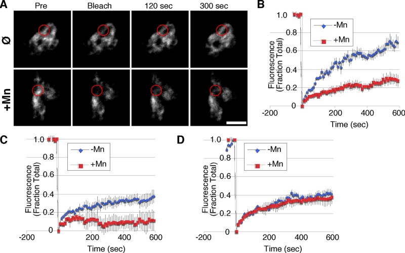
FIGURE 5:
Mn slows diffusion of GPP130 in the Golgi. (A) Fluorescence images of GPP130 tagged with GFP in untreated or Mn-treated cells immediately before a small zone of the Golgi was bleached and 0, 2, and 5 min after the bleaching. Circles indicate the position and average apparent size of the bleaching. Bar, 5 μm. (B) Quantified fluorescence levels of GPP130-GFP in the bleached zone at time points before and after bleaching for untreated and Mn-treated cells (n ≥ 10, ±SEM). (C) Quantified fluorescence levels of the chimeric GP73-GPP130 construct containing the wild-type GPP130 segment 36–175 in the bleached zone at time points before and after bleaching for untreated and Mn-treated cells (n ≥ 10, ±SEM). (D) Quantified fluorescence levels of the chimeric GP73-GPP130 construct containing the 88AAAA91 substitution in the GPP130 segment 36–175 in the bleached zone at time points before and after bleaching for untreated and Mn-treated cells (n ≥ 10, ±SEM).

