
FIGURE 1:
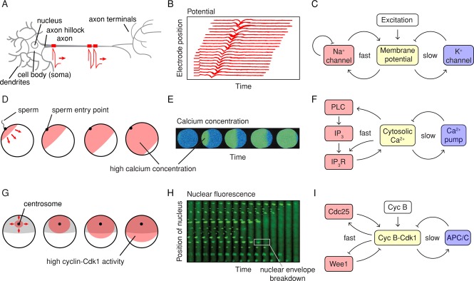
Examples of biological trigger waves. (A–C) Action potentials. (A) Action potentials are generated at the axon hillock and propagate distally down the axon. (B) Recordings of an action potential traveling down an axon, measured by an array of extracellular electrodes. The inward flux of Na+ during an action potential registers as a negative deflection of the potential registered by the extracellular electrodes. (Adapted from Bakkum et al., 2013.) (C) Schematic view of the circuit that generates the action potential. (D–F) Calcium waves in fertilized eggs. (D) Calcium waves are generated at the sperm entry point and spread across the egg. (E) Calcium concentrations as a function of time in a fertilized oocyte from the milky ribbon worm, Cerebratulus lacteus, as measured by ratiometric imaging after calcium green loading. (Taken from Stricker, 1999.) (F) Schematic view of the circuit that generates calcium waves. (G–I) Mitotic waves in Xenopus eggs. (G) About 1 h after fertilization and the postfertilization calcium wave, a wave of Cdk1 activation spreads from near the centrosome to the cortex of the cell. (H) Waves of mitosis in Xenopus egg extracts. Thin Teflon tubes were filled with cycling Xenopus egg extracts together with sperm chromatin and a nuclear localization signal–green fluorescent protein marker. Waves of nuclear envelope breakdown spread from the fastest regions of the cytoplasm, near the middle of this section of the tube, outward. (Taken from Chang and Ferrell, 2013.) (I) Schematic view of the circuit that generates waves of cyclin B-Cdk1 activation.