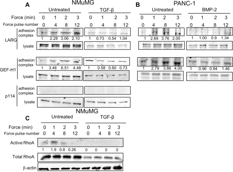
FIGURE 5:

LARG and GEF-H1 recruitment to adhesion complex during force decrease during TGF-β–induced EMT. (A, B) Effect of EMT on RhoA GEF recruitment in either NMuMG (A) or PANC-1 (B) cells. Indicated cells were incubated for 30 min with FN-coated beads (Guilluy et al., 2011) and stimulated with a force regimen (50 pN; 5 s force, 10 s recovery) using a rotating permanent magnet for different amounts of time (Materials and Methods). After magnetic separation of the adhesion complex, both the lysate and adhesion complex fractions were analyzed using Western blots. Representative of four independent experiments. Associated quantification of amount of protein in adhesion complex (bead-to-lysate ratios), relative to untreated cells without force stimulation, is provided. (C) Effect of EMT on RhoA activation in NMuMG cells. Cells were stimulated with a force regimen using a rotating permanent magnet as in A. RhoA activity in lysates was determined as described (Guilluy et al., 2011). Representative of three experiments. Associated quantification of amount of protein in adhesion complex (bead-to-lysate ratio), relative to untreated cells without force stimulation, is provided.
