
FIGURE 3:
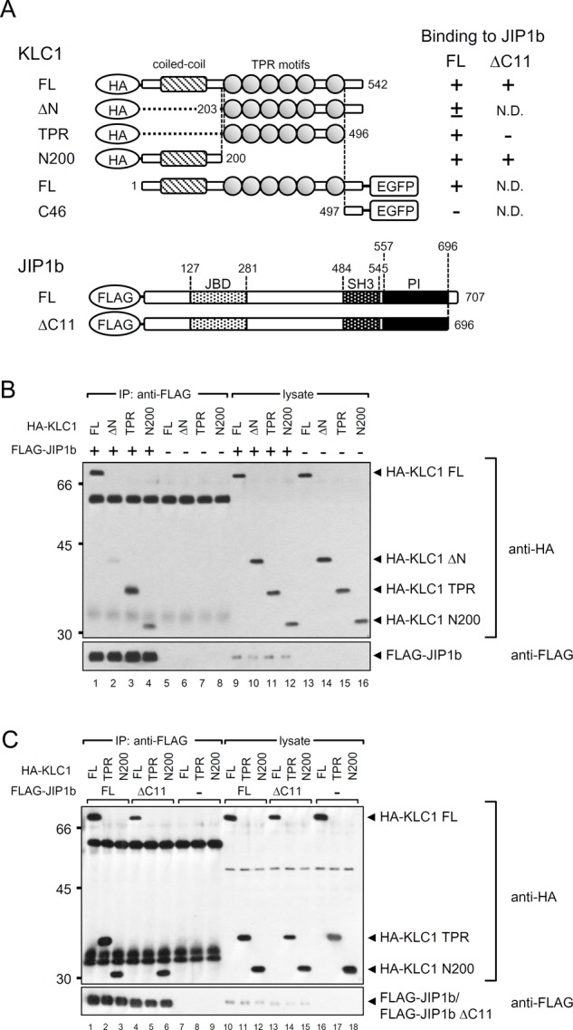
Interaction of the N-terminal half of KLC1 with JIP1b. (A) Structure of N-terminal HA- or C-terminal EGFP-tagged mouse KLC1 and N-terminal FLAG-tagged mouse JIP1b proteins. Numbers represent amino acid positions. The shaded box indicates the coiled-coil/heptad repeats region, and ovals indicate TPR motifs in KLC1. JIP1bΔC11 is the truncated protein lacking 11 C-terminal amino acids (697YTCPTEDIYLE707). JBD, JNK-binding motif; SH3, Src homology domain 3; PI, phosphotyrosine interaction or phosphotyrosine binding (PTB) domain. Summary of KLC1 interactions with JIP1b is shown on the right, classified as binding (+), weak binding (±), and nonbinding (–) in a coimmunoprecipitation assay. N.D., not determined. (B, C) Coimmunoprecipitation of KLC1 and JIP1b. The IPs and lysate samples were analyzed by immunoblotting with anti-HA and anti-FLAG antibodies. Numbers shown at the left indicate molecular weight standards (kilodaltons). Numbers shown below indicate lane numbers. Protein bands at ∼60 and 35 kDa in the IP lanes shown in this and later figures are derived from the antibody used.