FIGURE 3.

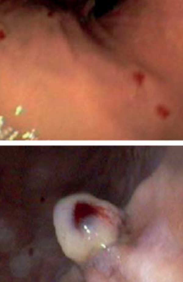
Upper endoscopy photos: Presence of multiple, friable, shiny telangiectasias in the fornix and gastric body, and tiny angiectases in the second duodenal portion

Upper endoscopy photos: Presence of multiple, friable, shiny telangiectasias in the fornix and gastric body, and tiny angiectases in the second duodenal portion