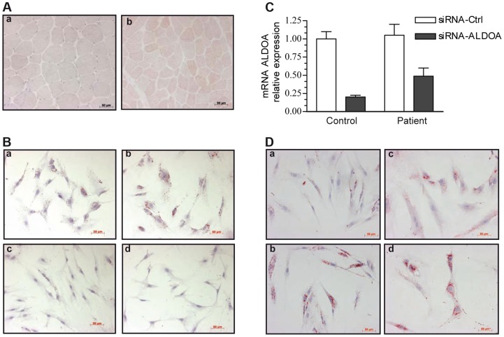
Figure 2. Oil-red-O staining of skeletal muscle and myoblasts from our patient and a control.
Images were taken with x20 magnification. 2A: Transverse cross-section of a left deltoid muscle biopsy of the patient shows the presence of numerous LDs, mainly in type 1 fibers, a: control, b: patient. 2B: Cytological oil-Red-O analysis of the patient myoblasts cultivated under basal (a) or pro-inflammatory conditions (TNFα+IL-1β) (b). LDs appear as red circular vacuoles in the cytoplasm. Treatment with dexamethasone alone (d) or combined with TNFα+IL-1β (c) reversed the LD phenotype. 2C: Relative knockdown of aldolase A expression in control and patient myoblasts. 2D: representative oil-red-O staining of mock-transfected control (a) and patient (c) cells, or aldolase A-siRNA-transfected control (b)and patient (d) myoblasts.

