
Figure 1. Membrane-intact mechanically loaded zebrafish myocytes.
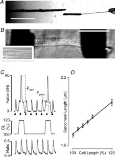
A, carbon rods coated with MyoTak glue were used to attach the myocytes: one rod was stiff (left; diameter 30 μm), and the other was compliant (right; diameter 10 μm). The latter was attached to a short stretch (30 μm) of the thicker probe, which increased attachment success. (Scale bar: 500 μm.) B, example of an attached membrane-intact enzymatically isolated myocyte. The inset shows a magnified region of the myocyte illustrating the appearance of sarcomere striations (observed in ∼30% of cells). (Scale bar: 50 μm.) C, example of an original recording of twitch force (top) and Indo-1 fluorescence ratio (bottom). Cells were stimulated at 1 Hz (arrowheads; 40 V, 2 ms). Every fifth twitch, cells were stretched (here: 20%). Increased cell length (CL) induced an increase in passive force as well as active developed twitch force. D, relationship between video FFT-derived diastolic sarcomere length and overall CL (n = 39).