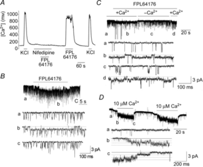
Figure 4. Activation of the 20 pS channel by FPL64176 and Ca2+.

A, tracing showing changes in [Ca2+]i in response to 20 mm KCl and 1 μm FPL64176 in the presence and absence of 1 μm nifedipine in isolated rat glomus cells. B, perfusion of a cell-attached patch showing TASK openings (tracing a) with 1 μm FPL64176 activated the 20 pS channel (tracing b). Washout of the agonist closed the 20 pS channel (tracing c). Pipette potential was 0 mV. C, application of FPL64176 (1 μm) to a cell-attached patch activated the 20 pS channel only when Ca2+ was present in the bath solution (tracings b and d). Perfusion with Ca2+-free solution closed the channel (tracing c). Pipette potential was 0 mV. D, Ca2+ applied to inside-out patch activated the 20 pS channel reversibly.
