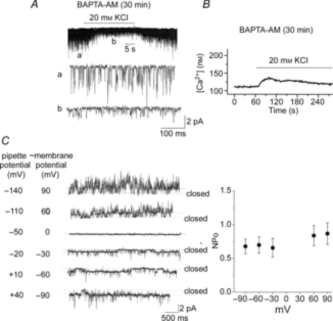
Figure 5. Lack of voltage dependence of the 20 pS channel.

A, glomus cells were incubated with 10 μm BAPTA-AM for 30 min. Current tracings show TASK openings from cell-attached patches with pipette solution containing 140 mm KCl and externally perfused with physiological solution containing 5 mm KCl first, then 20 mm KCl and then back to 5 mm KCl. B, glomus cells were incubated with 10 μm BAPTA-AM for 30 min, and changes in [Ca2+] were measured in response to 20 mm KCl. An averaged tracing from 28 cells is shown. C, cell-attached patches were formed on rat glomus cells with pipette solution containing 140 mm KCl and externally perfused with physiological solution. FPL64176 (1 μm) was applied to the bath to activate the 20 pS channel. Current tracings are shown at various pipette potentials. In this particular patch, the current reversed at −50 mV pipette potential (membrane potential of ∼0 mV). In four other patches, the current reversed at pipette potential between −60 and −40 mV. On the right, the graph plots the 20 pS channel activity as a function of the adjusted cell membrane potential. Each point is the mean ± SD from five patches. No significant differences were present between data points (P > 0.05).
