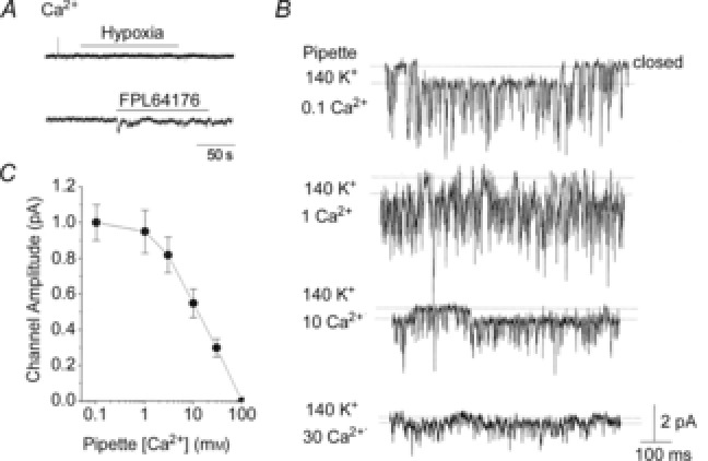
Figure 7. Inhibition of the 20 pS channel conductance by extracellular Ca2+.

A, cell-attached patches were formed on rat glomus cells with pipette containing 100 mm CaCl2. In physiological bath solution, no channels were activated by hypoxia or FPL64176. The current tracings shown were obtained when the pipette potential was 0 mV. No channels were present with pipette potentials ranging from −120 to +100 mV. B, cell-attached patches were formed with varying levels of Ca2+ in the pipette together with 140 mm KCl. FPL64176 was added to the bath solution to activate the 20 pS channel. Current tracings were obtained at pipette potential set at 0 mV. C, amplitude of the 20 pS channel plotted as a function [Ca2+] in the pipette. Each point is the mean ± SD (n = 5).
