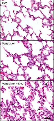
Figure 6. Representative H&E-stained sections of the lung.

Sections of the right upper lung lobe of unventilated controls (UVC), ventilated lambs and lambs that were ventilated and received EPO (Ventilation + EPO). Note the increase in inflammation and alveolar wall thickness after ventilation, which is further exacerbated by EPO administration. Arrows indicate accumulation of red blood cells within the airways. Arrowhead indicates increased accumulation of inflammatory cells within the interstitium.
