FIGURE 1:

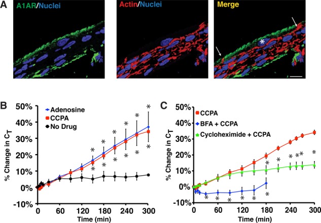
A1AR-stimulated exocytosis is dependent on protein synthesis and secretion. (A) Cryosection of rat bladder epithelium labeled with an A1AR-specific antibody (green), rhodamine–phalloidin to label the actin cytoskeleton (red), and TOPRO-3 (blue) to label the nucleus. Right, merge. An umbrella cell is marked with an asterisk and the white arrows mark the apicolateral borders of the cell. Scale bar, 12 μm. (B) Rabbit uroepithelium was mounted in Ussing chambers and after equilibration left untreated (no drug), or 1 μM adenosine or 500 nM CCPA was added to the mucosal hemichamber. Percentage changes in CT were monitored. (C) Rabbit tissue was left untreated, pretreated with 5 μg/ml brefeldin A (BFA) for 30 min, or pretreated with 100 ng/ml cyclohexamide for 60 min. CCPA (500 nM) was then added to the mucosal hemichamber and CT recorded. Data for CCPA alone are reproduced from B. (B, C) Mean changes in CT ± SEM (n ≥ 4). Significant differences (p < 0.05), relative to no drug in A or CCPA in B, are indicated with an asterisk.
