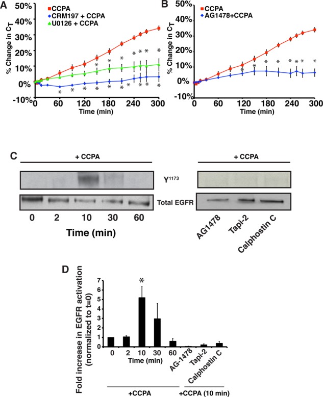
FIGURE 2:

The A1AR transactivates the EGF receptor. (A) The mucosal surface of rabbit uroepithelium was pretreated with 25 ng/ml CRM197 for 25 min or with 10 μM U0126 for 60 min. CCPA (500 nM) was then added to the mucosal hemichamber, and CT was recorded. (B) Rabbit uroepithelium was pretreated with 1 μM AG1478 for 30 min, and then CCPA (500 nM) was added to the mucosal hemichamber and CT was recorded. (A, B) CCPA control data are reproduced from Figure 1B. Mean changes in CT ± SEM (n ≥ 3). Statistically significant differences (p < 0.05), relative to CCPA treatment alone, are marked with an asterisk. (C, D) Rabbit uroepithelium was either left untreated (left) or treated with AG1478 (1 μM) for 25 min, Tapi-2 (15 μM) for 90 min, or calphostin C (500 nM) for 60 min (right). CCPA (500 nM) was then added to the mucosal hemichamber. Left, cells were lysed at the indicated time points. Right, cells were lysed at the 10-min time point. Equal amounts of proteins were resolved by SDS–PAGE and immunoblots probed with a rabbit anti–EGFR-phospho-Y1173 antibody or rabbit anti-EGFR antibody. (D) Quantification of Y1173 phosphorylation. Data (mean ± SEM, n ≥ 3) are reported as fold increase above untreated tissue samples at t = 0. Statistically significant values (p < 0.05) above t = 0 are marked by an asterisk.
