
FIGURE 3:
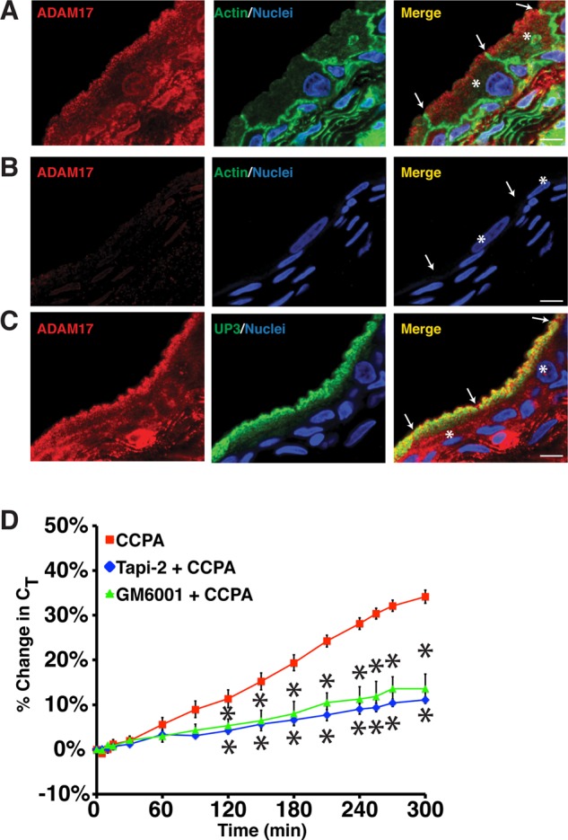
ADAM17 expression in the uroepithelium. (A–C) Cryosections of rat bladder uroepithelium were reacted with antibodies specific for ADAM17 (A), a mixture of antibody and inhibitory peptide (B), or antibodies specific for ADAM17 and uroplakin 3a (C). After incubation with fluorophore-labeled secondary antibodies, the samples were analyzed using confocal microscopy. Where indicated, actin was labeled with phalloidin and nuclei were labeled with TOPRO-3. The umbrella cells are marked with the asterisk in the merged images and the apicolateral junction is indicated by arrowheads; scale bar, 10 μm. (D) Rabbit uroepithelium was pretreated with Tapi-2 (15 μM) or GM6001 (15 μM) for 90 min, CCPA (500 nM) was added, and CT was recorded. Control CCPA data are reproduced from Figure 1B. Data are mean ±SEM (n ≥ 3), and values significantly different (p < 0.05) from CCPA alone are marked with an asterisk.