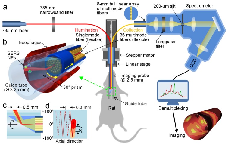
Fig. 1.
Schematic of a customized spectral-imaging endoscope and SERS-endoscopy system. (a) Experimental platform to detect multiplexed NPs within the esophagus of a rat. (b) Zoom-in rendering of the prism and fiber-bundle imaging probe within a glass guide tube and rat esophagus (Media 1 (3.1MB, AVI) ). (c) Cross-sectional illustration of the illumination and collection beam paths. (d) Scanning trajectory for comprehensive imaging of the rat esophagus. To illustrate our scanning strategy, the guide tube is unfolded and the neutral position (0°) and turning points ( ± 180°) are marked. The probe rotates between ± 180° as it is slowly pulled out of the esophagus. The colored circles represent the spot size of the laser illumination and the spacing between consecutive spectral acquisitions (image pixels).

